Looking at a heart
Class practical or demonstration
It is possible to learn about the function of the heart from diagrams, models and animations. However, the experience of dissecting real animal material adds an extra dimension to understanding the structure of the heart and the relationship of structure to function. The texture and thickness of vessel and chamber walls, and the movement of different kind of valves are unforgettable.
Lesson organisation
This will depend on how many hearts you obtain and on how many of your students opt out of handling (or closely observing) the specimens.
If you have access to a magnifying video camera you could project a demonstration dissection. If you have a plastic model heart, a number of students could work from that. Prepare a handout with a drawing of the heart and questions for students who are not taking a practical part in the activity. Students who are unwilling to handle hearts, but keen to see what is going on could take digital photographs of the heart in different stages of dissection.
Allow 5-10 minutes for exploring the heart and attached vessels before starting to cut into the material.
Apparatus and Chemicals
For each group of students:
Heart, from butcher/ supplier, Note 2
Tray
Scalpel
Dissecting scissors
Ruler, 15 cm, that can be disinfected after use
Access to thin vinyl or nitrile gloves
Access to warm water and soap for handwashing
For the class – set up by technician/ teacher:
Disinfectant, to clean bench tops and rulers at end of procedure, Note 1
Health & Safety and Technical notes
Wear eye protection whenever there is a risk to the eyes, for example, when changing scalpel blades, cutting cartilage or when the dissection material has been preserved.
1 Disinfectant: Use Virkon, following manufacturer’s instructions.
2 Sometimes butchers cut the blood vessels off the top of the heart. They may also remove the atria and cut into the ventricles at the abbatoir (as part of the inspection process). If you discuss your needs with your butcher, they may be able to limit this, and provide hearts in the form that you need. If you order a ‘pluck’ – heart and lungs together – you usually get long lengths of blood vessel still attached to the heart. You can cut sections of these veins and arteries for studies of vascular structure – see Elastic recoil in arteries and veins. And you can freeze the lungs for later study.
3 Safety of handling butcher material: Anything that a butcher sells must have been passed as ‘fit for human consumption’, but may be carrying food-poisoning bacteria. We don’t wear gloves when preparing meat in a kitchen, so it is not necessary to wear gloves, but it is necessary to wash hands thoroughly after handling the material and before leaving the laboratory. The government has enacted the Specified Risk Materials (SRM) Regulations and these determine the parts of slaughtered animals which may be capable of transmitting spongiform encephalopathies and therefore must not be supplied. Only materials from cattle, sheep and goats are currently specified and only under certain circumstances. Thus anything that you can obtain through normal channels should not be SRM and could therefore be used. Do not store the material used for dissection in refrigerators used for food.
4 Cleaning equipment and work surfaces: Dissecting instruments are best autoclaved after use because most disinfectants attack metal instruments. Clean contaminated equipment such as dissecting boards with hot water and detergent and soak for at least 10 minutes in a freshly-prepared 1% solution of Virkon disinfectant. Wipe down working surfaces with 1% Virkon, and leave wet for 10 minutes before drying off. If you perform dissections on layers of newspaper you may protect the bench surface enough for it not to need to be disinfected.
5 If students receive cuts or scratches during dissection, wash the wound in cold running water. Allow minor wounds to bleed freely and refer to a first aider.
Ethical issues
The animal material used has not been produced for the purpose of the investigation – it is a regular by-product of the meat industry, available for consumption from some butchers, or part of the waste stream if not used for dissection.
Some students may be sensitive to the use of any animal product in this way, or to the very existence of animals bred and raised for human consumption.
Some students may have religious or cultural objections to handling dead animal material, particularly pigs or cows, and depending on the method of slaughter.
Procedure
SAFETY: Take care with sharp dissecting tools.
Observe careful hygiene precautions after the dissection.
Preparation
a Purchase hearts from butcher or other supplier, after discussing how the hearts are to be used. Note 1.
b Ensure all dissecting tools are clean and sharp.
c Decide how the hearts are to be disposed of. This should either be by incineration, or in a dustbin or container used for food wastes that are not to be fed to animals.
d Ensure first aid kit for cuts is available in the teaching room.
Investigation 1 Looking at the outside of the heart
e Note the general shape and size of the heart.
f Measure its size and mass. Estimate its external volume.
g Identify the vessels entering and leaving the heart. Arteries have thick, rubbery walls. Veins have much thinner walls. Feel inside these vessels with your fingers and feel the texture and strength of both vessels. Try to describe what you feel.
h Look inside the main arteries and veins. If you see any structures attached to the walls, try to decide what they are, what they might do, and how they might work.
i Identify the atria and ventricles. Try to describe any differences in the structures of the walls of the atria and ventricles.
j Decide which you think is the right side of the heart and which is the left. What has helped you to decide?
k Examine the surface of the heart for blood vessels.
l Note the colour and texture of the different parts of the heart.
Investigation 2 The internal structure of the heart
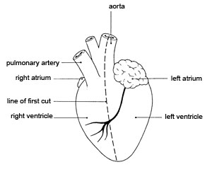
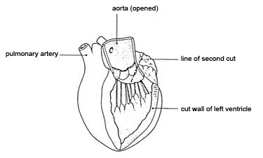
m Look at the left-hand diagram. Make a long cut down through the aorta and the left ventricle to the tip of the heart (‘apex’), as shown in the diagram. The position of the blood vessels on the surface will help you to make this cut in the right place. Identify the coronary artery that supplies the heart muscle itself with blood.
n Pull the edges of the ventricle apart and examine the inside of the ventricle and the aorta. At the base of the aorta, you will see the structures mentioned in h. Examine them carefully.
o Look at the right-hand diagram. Cut carefully upwards into the left atrium along the line shown in the diagram.
p Measure and record the thickness of the walls of the atrium and the ventricle.
q Examine the right side of the heart in a similar way.
r Look at the areas where an atrium joins a ventricle. Examine the structures there. These are valves separating the chambers of the heart. You should see flaps of thin tissue, with tough ‘threads’ attached to the base of the flaps. Count how many threads there are on each side of the heart. Think about how these valves might work.
Downloads
Download the student sheet ![]() Looking at a heart (78 KB) with questions and answers.
Looking at a heart (78 KB) with questions and answers.
Health & Safety checked, December 2008
Web links
http://www.heartworks.me.uk/index.php?page=videos&lang=uk
This project has developed software that shows an animated 3-D heart, beating steadily, alongside ultrasound images and an ECG. It is intended as an interactive tool for teaching medical students, so much of the information on the site is beyond the scope of students at school. However, the clips available give a really nice sense of a beating heart. Go to the link above, and see a sample of the output.
http://www.nhlbi.nih.gov/health/dci/Diseases/hhw/hhw_pumping.html
This is a short 3-D animation of the heart in action, showing the detailed movement of the valve flaps and the heart muscle. The commentary has an American accent, but is quite clear.
(Websites accessed October 2011)